Featured Publications
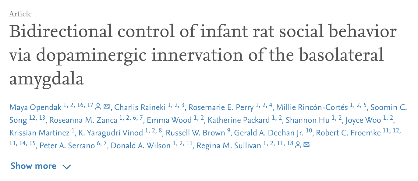
Social interaction deficits seen in psychiatric disorders emerge in early-life and are most closely linked to aberrant neural circuit function. Due to technical limitations, we have limited understanding of how typical versus pathological social behavior circuits develop. Using a suite of invasive procedures in awake, behaving infant rats, including optogenetics, microdialysis, and microinfusions, we dissected the circuits controlling the gradual increase in social behavior deficits following two complementary procedures—naturalistic harsh maternal care and repeated shock alone or with an anesthetized mother. Whether the mother was the source of the adversity (naturalistic Scarcity-Adversity) or merely present during the adversity (repeated shock with mom), both conditions elevated basolateral amygdala (BLA) dopamine, which was necessary and sufficient in initiating social behavior pathology. This did not occur when pups experienced adversity alone. These data highlight the unique impact of social adversity as causal in producing mesolimbic dopamine circuit dysfunc-tion and aberrant social behavior.
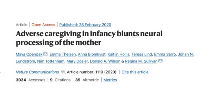
The roots of psychopathology frequently take shape during infancy in the context of parent-infant interactions and adversity. Yet, neurobiological mechanisms linking these processes during infancy remain elusive. Here, using responses to attachment figures among infants who experienced adversity as a benchmark, we assessed rat pup cortical local field potentials (LFPs) and behaviors exposed to adversity in response to maternal rough and nurturing handling by examining its impact on pup separation-reunion with the mother. We show that during adversity, pup cortical LFP dynamic range decreased during nurturing maternal behaviors, but was minimally impacted by rough handling. During reunion, adversity-experiencing pups showed aberrant interactions with mother and blunted cortical LFP. Blocking pup stress hormone during either adversity or reunion restored typical behavior, LFP power, and cross-frequency coupling. This translational approach suggests adversity-rearing produces a stress-induced aberrant neurobehavioral processing of the mother, which can be used as an early biomarker of later-life pathology.
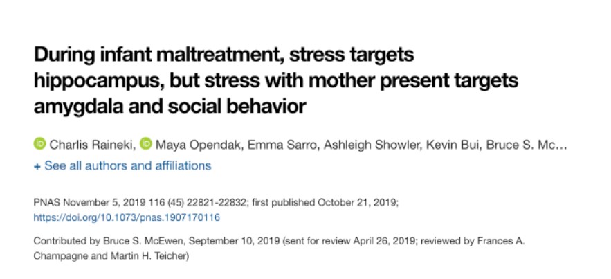
Infant maltreatment increases vulnerability to physical and mental disorders, yet specific mechanisms embedded within this complex infant experience that induce this vulnerability remain elusive. To define critical features of maltreatment-induced vulnerability, rat pups were reared from postnatal day 8 (PN8) with a maltreating mother, which produced amygdala and hippocampal deficits and decreased social behavior at PN13. Next, we deconstructed the maltreatment experience to reveal sufficient and necessary conditions to induce this phenotype. Social behavior and amygdala deficits (volume, neurogenesis, c-Fos, local field potential) required combined chronic high corticosterone and maternal presence (not maternal behavior). Hippocampal deficits were induced by chronic high corticosterone regardless of social context. Causation was shown by blocking corticosterone during maltreatment and sup- pressing amygdala activity during social behavior testing. These results highlight (1) that early life maltreatment initiates multiple pathways to pathology, each with distinct causal mechanisms and outcomes, and (2) the importance of social presence on brain development.
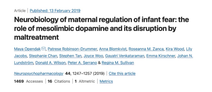
Child development research highlights caregiver regulation of infant physiology and behavior as a key feature of early life attachment, although mechanisms for maternal control of infant neural circuits remain elusive. Here we explored the neurobiology of maternal regulation of infant fear using neural network and molecular levels of analysis in a rodent model. Previous research has shown maternal suppression of amygdala-dependent fear learning during a sensitive period. Here we characterize changes in neural networks engaged during maternal regulation and the transition to infant self-regulation. Metabolic mapping of 2-deoxyglucose uptake during odor-shock conditioning in postnatal day (PN)14 rat pups showed that maternal presence blocked fear learning, disengaged mesolimbic circuitry, basolateral amygdala (BLA), and plasticity-related AMPA receptor subunit trafficking. At PN18, when maternal presence only socially buffers threat learning (similar to social modulation in adults), maternal presence failed to disengage the mesolimbic dopaminergic system, and failed to disengage both the BLA and plasticity-related AMPA receptor subunit trafficking. Further, maternal presence failed to block threat learning at PN14 pups following abuse, and mesolimbic dopamine engagement and AMPA were not significantly altered by maternal presence—analogous to compromised maternal regulation of children in abusive relationships. Our results highlight three key features of maternal regulation: (1) maternal presence blocks fear learning and amygdala plasticity through age-dependent suppression of amygdala AMPA receptor subunit trafficking, (2) maternal presence suppresses engagement of brain regions within the mesolimbic dopamine circuit, and (3) early-life abuse compromises network and molecular biomarkers of maternal regulation, suggesting reduced social scaffolding of the brain.


